Application note ·
Cathode-material recycling and battery pilot lines need sub-micron CT, not just lab analytics
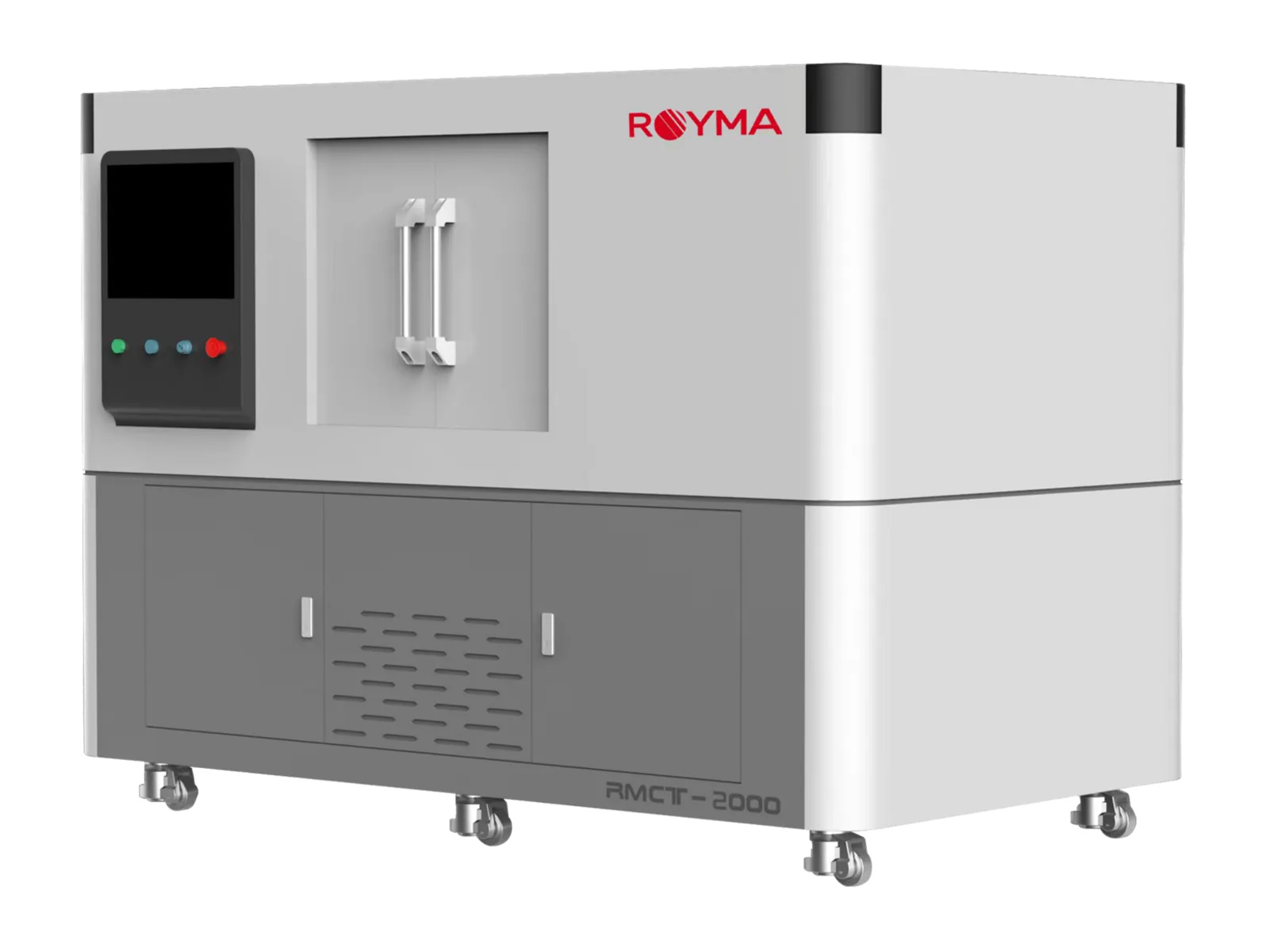
Royma Industrial CT offers sub-micron computed tomography (CT) for battery materials with the RMCT2000HR nanofocus machine. It resolves detail down to 0.5 µm in 2D and 1 µm in 3D, so recycling plants and battery pilot lines see the inside of recovered powders, reformed cathode material and electrode samples without cutting them open.
The application note is written for process engineers and laboratory managers who qualify battery materials. It covers the inspection task, the choice between the RMCT2000 and the RMCT2000HR, and what a scan shows.
Sub-micron CT adds the third dimension to materials analysis
Cathode-material recycling and battery research are growing across Europe. New recycling lines recover and re-form active material. Pilot lines test new cell chemistries and electrode formats at small scale before they move to production.
Both need to know whether a material is fit for use. Chemical analysis states what the material is made of. Particle-size measurement states how large the particles are. Neither shows how particles are built inside, or how they pack together in a coated electrode.
That structure decides how a recovered or new material behaves in a cell. Cross-sections and microscope images show it, but only on one cut plane, and the sample is lost. CT shows the complete volume of a sample in 3D and leaves it intact for further tests.
The inspection task: particles, pores and coatings
Three questions recur in recycling plants and pilot lines:
- Recovered and re-formed cathode powder: are the particles intact, or do they carry internal cracks, voids and inclusions from the recycling process?
- Coated electrodes: how are porosity and compaction distributed through the thickness of the coating, and where are denser or looser zones?
- Pilot-line samples: where do delamination, micro-cracks or foreign particles appear between coating, current collector and separator?
Each of these features sits in the micrometre range or below. A CT machine for this task therefore needs a very small focal spot, a short distance between source and sample, and a stable set-up over a long scan.
Nanofocus CT resolves the micro-structure of battery materials
The RMCT2000 and the RMCT2000HR are Royma’s laboratory CT machines for small, fine-structured samples. Both use an open-tube nanofocus X-ray source that runs from 110 to 190 kV.
The two versions differ in resolution. The RMCT2000 reaches 2.5 µm in 2D and 3 µm in 3D. The RMCT2000HR reaches 0.5 µm in 2D (JIMA) and 1 µm in 3D. For single particles and thin electrode samples, the RMCT2000HR is the right choice. For larger electrode stacks and component sections, where micrometre detail is enough, the RMCT2000 covers the task.
Both machines take samples up to 150 × 150 × 150 mm and 5 kg. Measurement accuracy is stated as 3.0 + L/50 µm, L being the measured length.
“Sub-micron CT shows a recovered cathode particle from the inside, not only its surface and its chemistry,” says Gabor Szabo, Sales Director Europe at Royma Europe. “You see cracks, voids and inclusions in 3D and keep the sample for your other tests.”
What the data shows
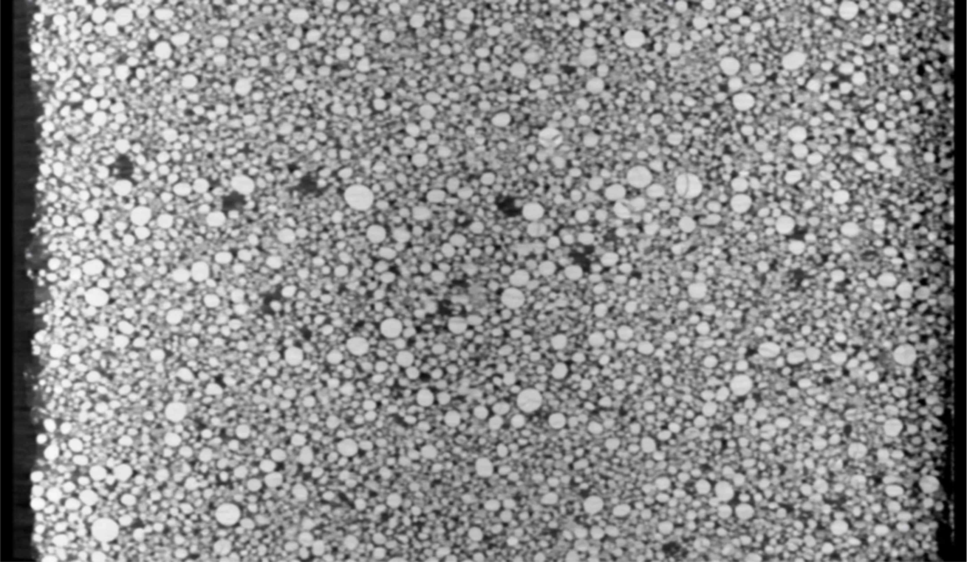
A nanofocus scan of a powder sample shows each particle as a volume. You can see internal cracks, hollow cores and inclusions, and count how often they occur across a sample. Recovered material can then be compared with virgin material on the same scale.
A scan of a coated electrode shows the pore network and the packing of the particles through the coating thickness. Porosity and compaction can be assessed layer by layer, instead of as a single average value for the whole electrode.
In pilot-line samples, the same volume shows micro-porosity, micro-voids and micro-cracks, as well as delamination between layers. Because the scan is non-destructive, a sample that shows a defect can go on to cross-sectioning or electrochemical testing afterwards. The CT result then tells you where to cut.
“Recycling and pilot lines change their process often,” Szabo adds. “A CT volume of each new batch gives your team a structural reference that stays comparable from run to run.”
Practical notes for sample preparation
The finest RMCT2000HR resolution is reached on small samples placed close to the source. For particle analysis, prepare a small, well-fixed powder sample so that particles do not move during the scan. For electrodes, cut a narrow strip or punch a small disc from the coated foil.
Plan the sample size around the resolution you need. Larger samples fit the machine up to 150 mm, but the voxel size grows with the field of view. Where both overview and detail matter, scan a larger section on the RMCT2000 first and a small sub-sample on the RMCT2000HR afterwards.
Material, particle size and coating thickness all affect the result. We therefore recommend a test scan of your own samples before a configuration is fixed. More typical tasks are described on the batteries and material analysis application pages.
| Stated figure | RMCT2000 | RMCT2000HR |
|---|---|---|
| Role | Laboratory nanofocus CT | Laboratory nanofocus CT |
| X-ray source | Open-tube nanofocus, 110–190 kV | Open-tube nanofocus, 110–190 kV |
| 2D resolution | 2.5 µm | 0.5 µm (JIMA) |
| 3D resolution | 3 µm | 1 µm |
| Sample size | Up to 150 × 150 × 150 mm | Up to 150 × 150 × 150 mm |
| Sample weight | Up to 5 kg | Up to 5 kg |
| Measurement accuracy | 3.0 + L/50 µm | 3.0 + L/50 µm |
Test scans of your battery materials
Royma Europe offers test scans of recovered cathode powder, reformed active material and electrode samples on the RMCT2000 and RMCT2000HR, with an evaluation of the resolution reached on your own material.
Contact: Gabor Szabo, Sales Director Europe, Royma Europe – gabor_szabo@roymatek.eu – +49 15156503316 – www.roymatek.eu
Royma Europe, Leibniz Universität Hannover – Institut für Produktentwicklung und Gerätebau, Gebäude 8143, An d. Universität 1, 30823 Garbsen, Germany.