
Applications
Batteries.
Cells, modules and packs.
Inspection tasks
Find the fault before formation.
Electrode alignment, weld quality and foreign particles decide whether a cell is safe. X-ray and CT show all three without opening the cell, in the laboratory and at the line.
Electrode overhang
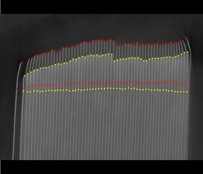
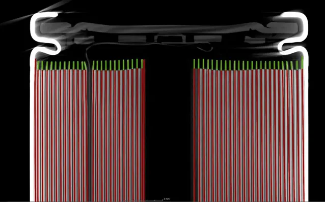
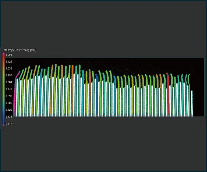
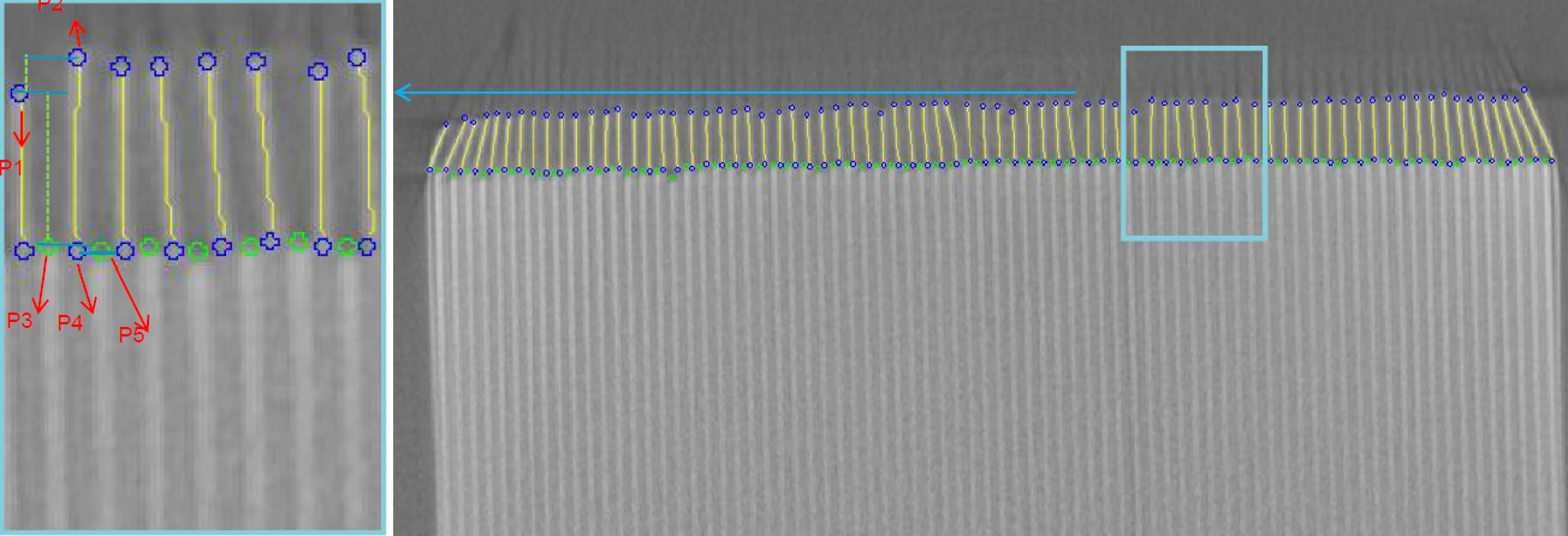
The anode must overlap the cathode on every layer. CT measures the overhang layer by layer in wound and stacked cells; 2D X-ray checks it at line speed.
Tabs, welds and housing
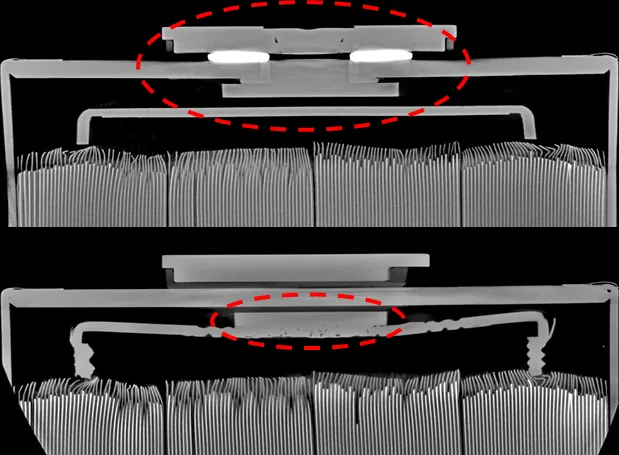
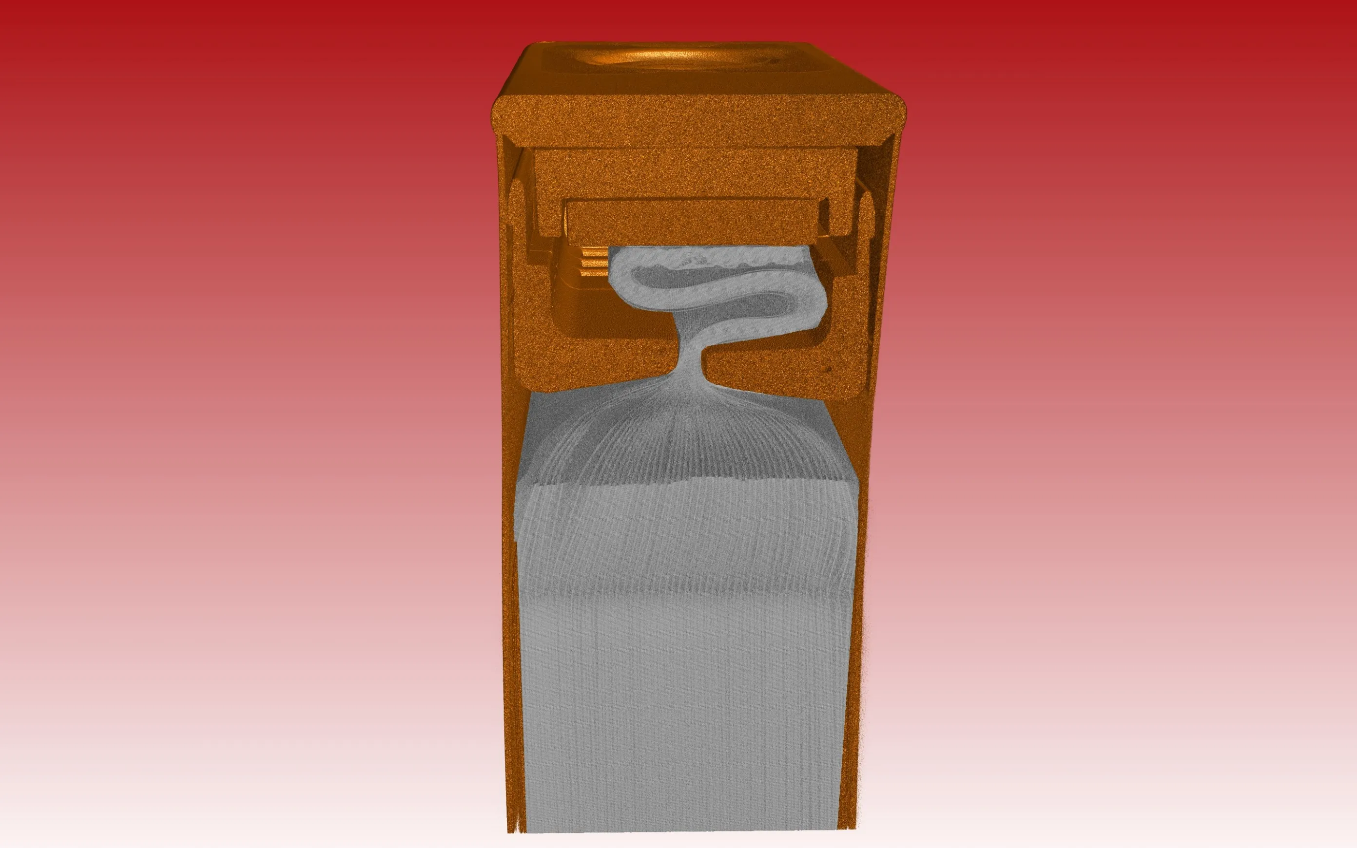
Folded tabs, weld defects and the distance between electrode and can are hidden behind the housing. A CT slice shows them in the assembled cell.
Foreign particles
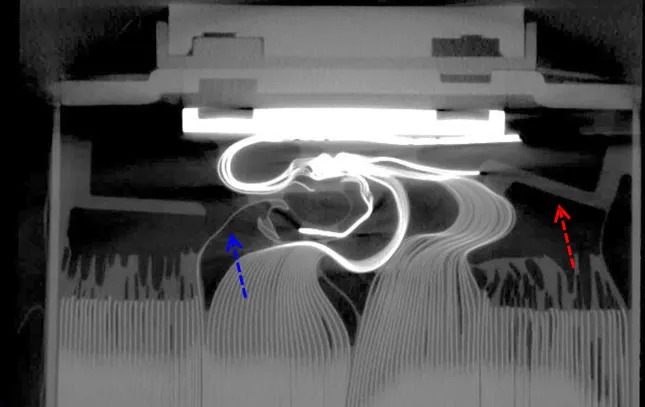
Metal particles in the cell can pierce the separator and cause an internal short circuit. CT locates each particle in three dimensions, so its size and its position between the layers are known.
All these inspection tasks are covered in detail in our batteries presentation.
Scan examples
What the data shows.
Systems
One technology from the laboratory to the line.
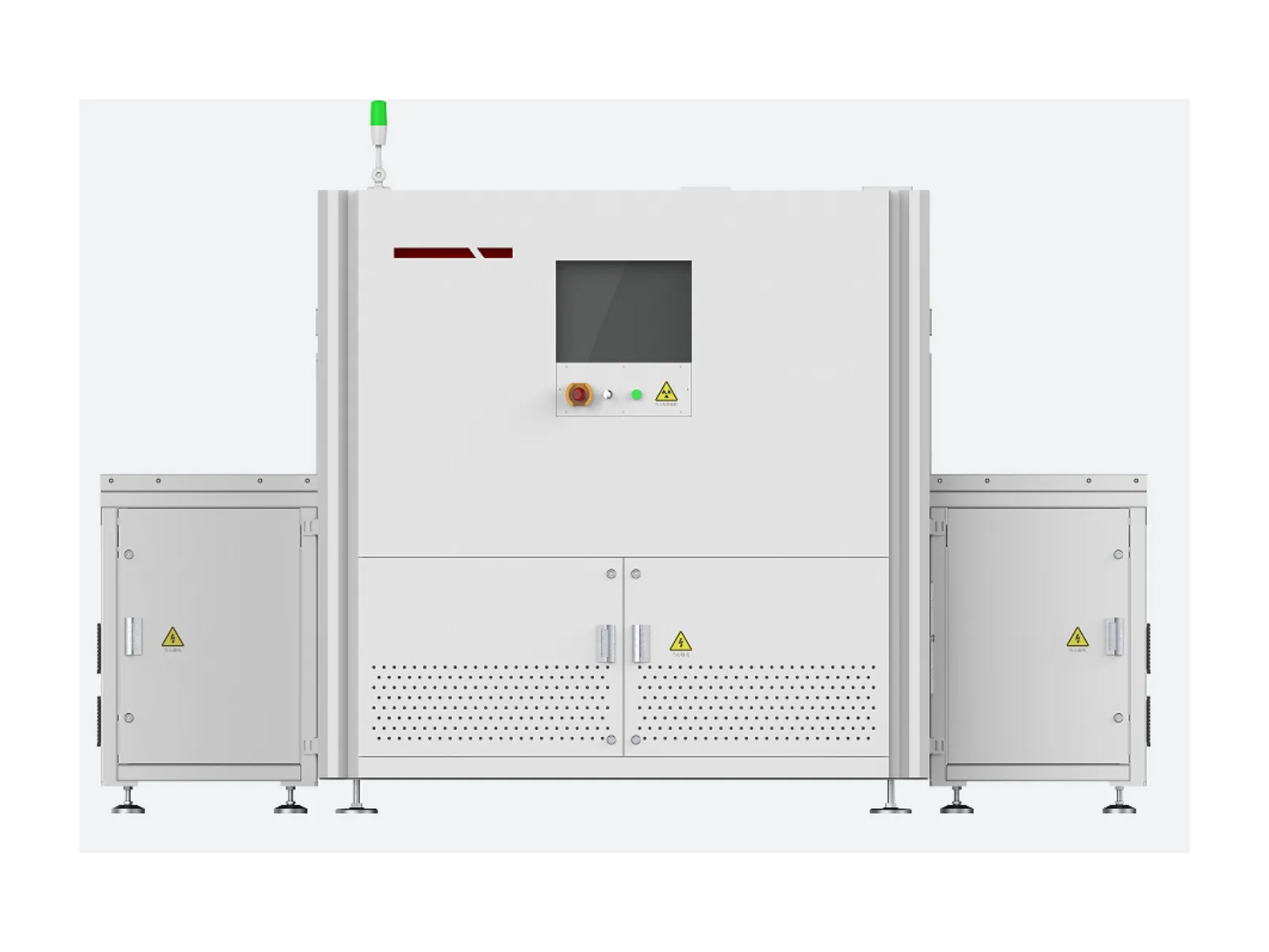
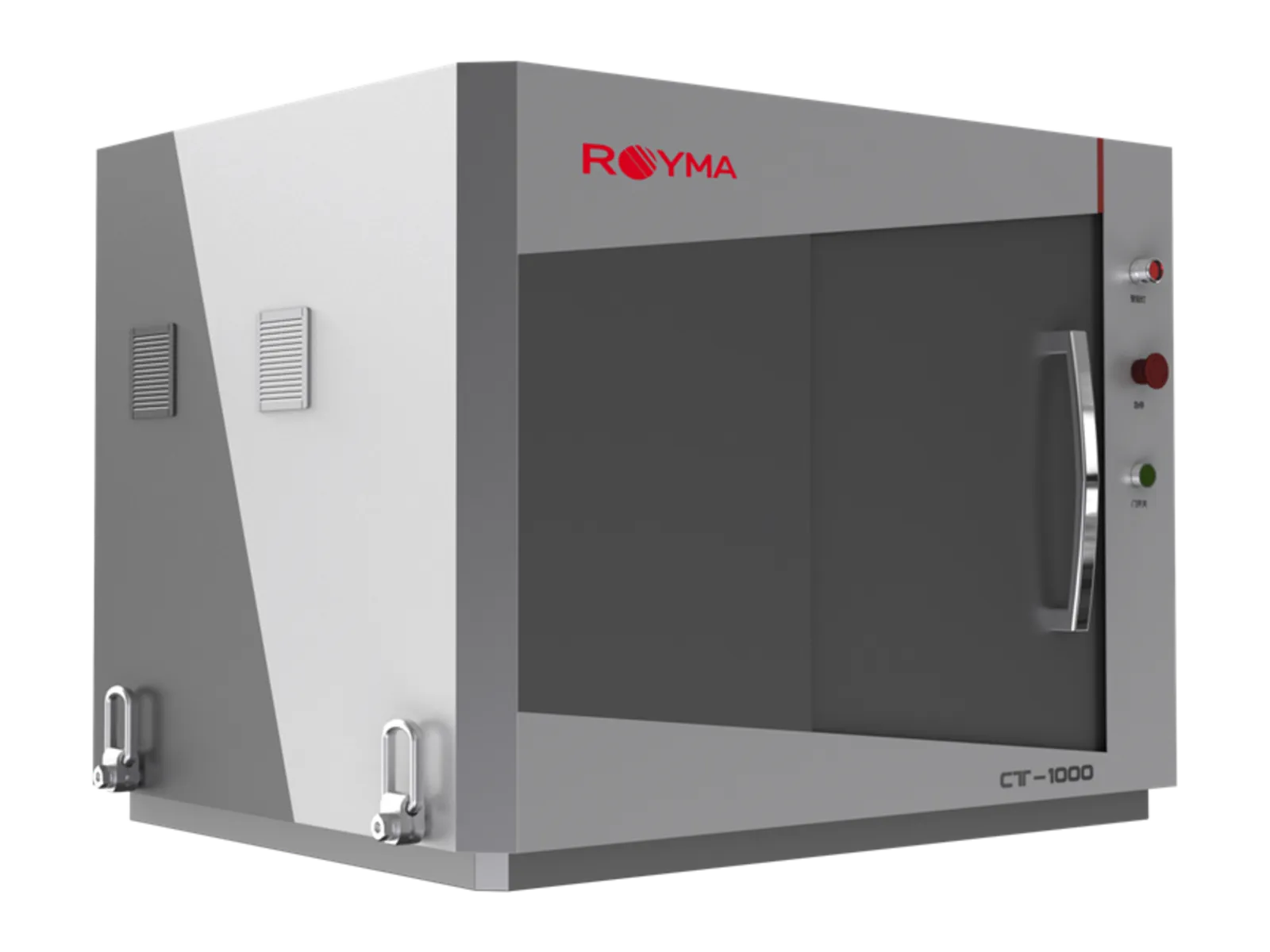
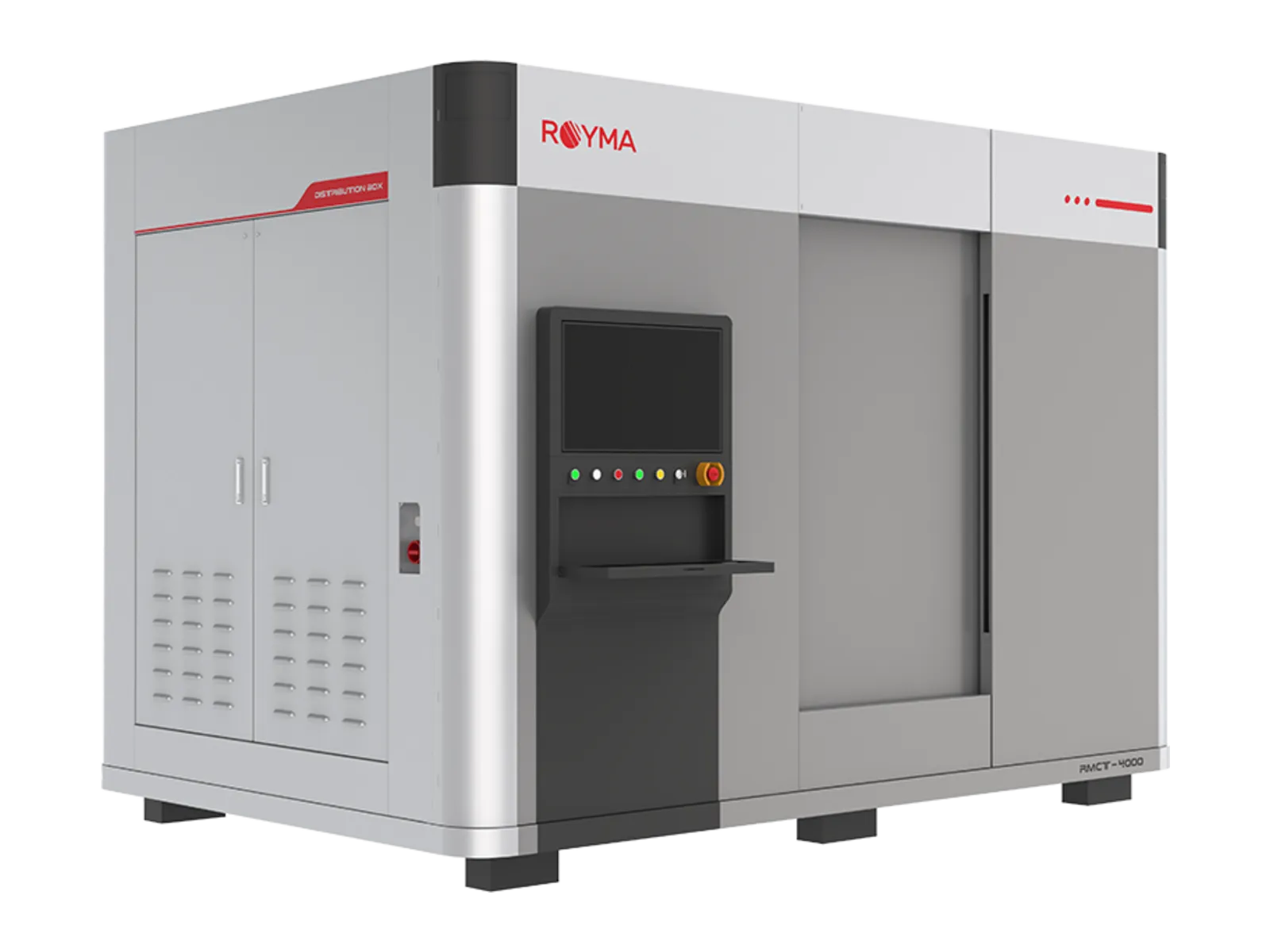
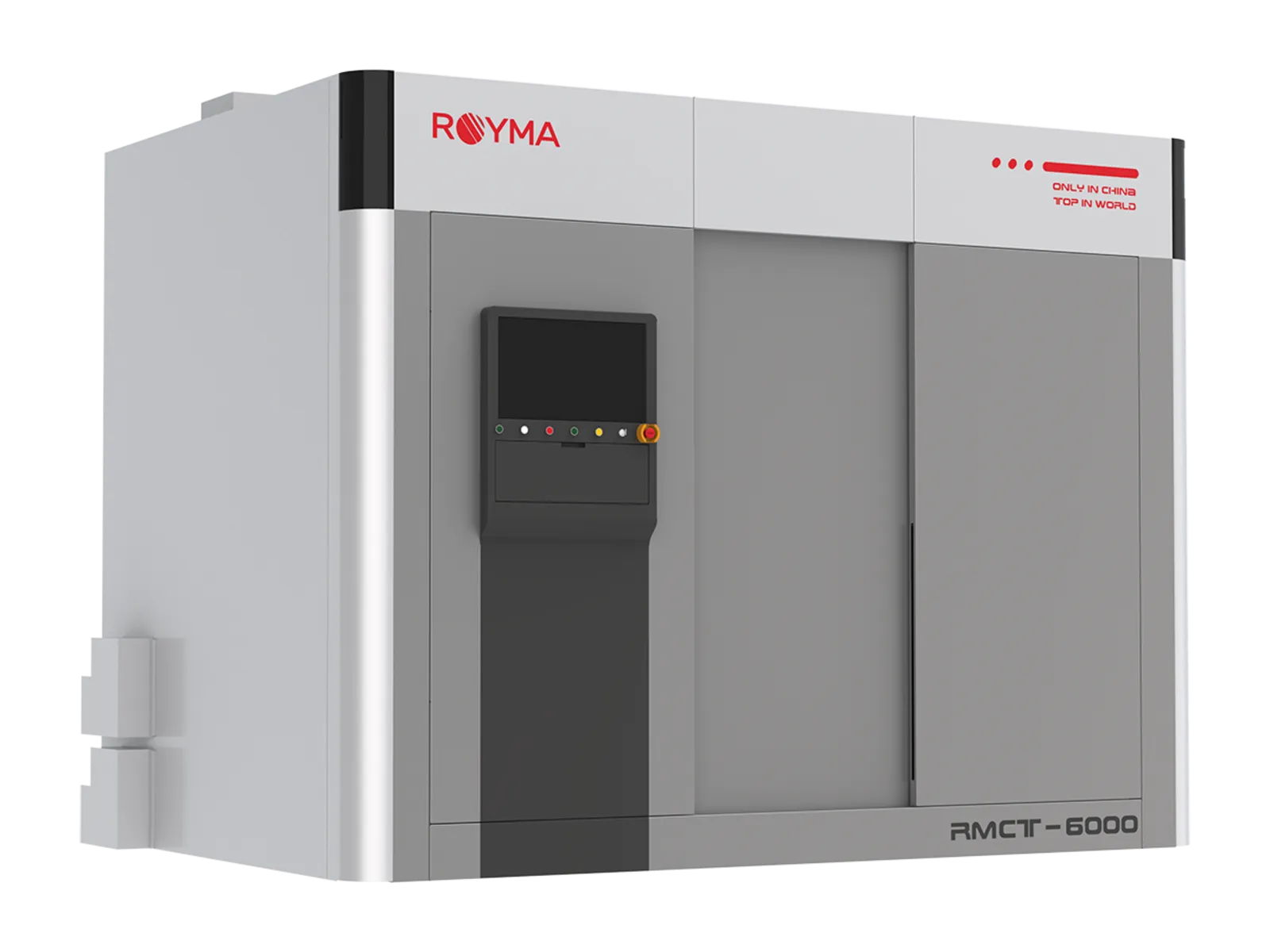
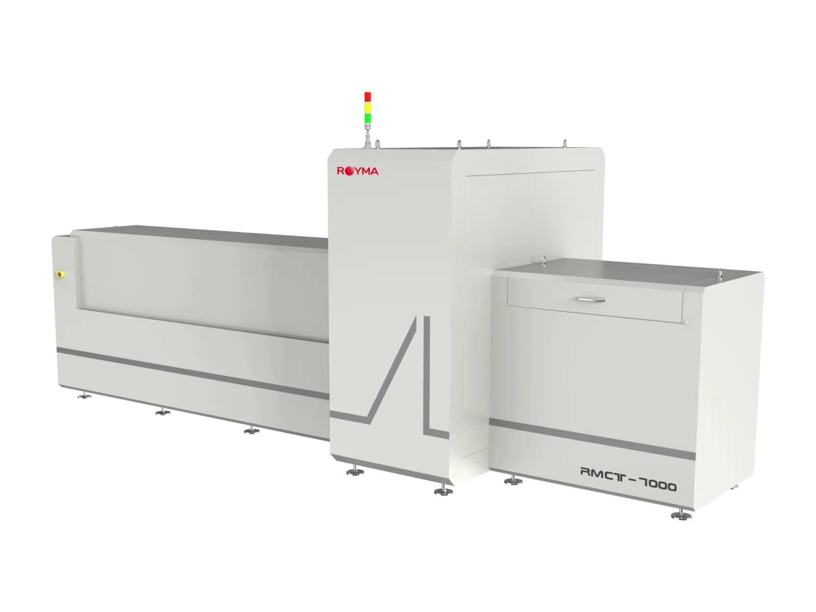
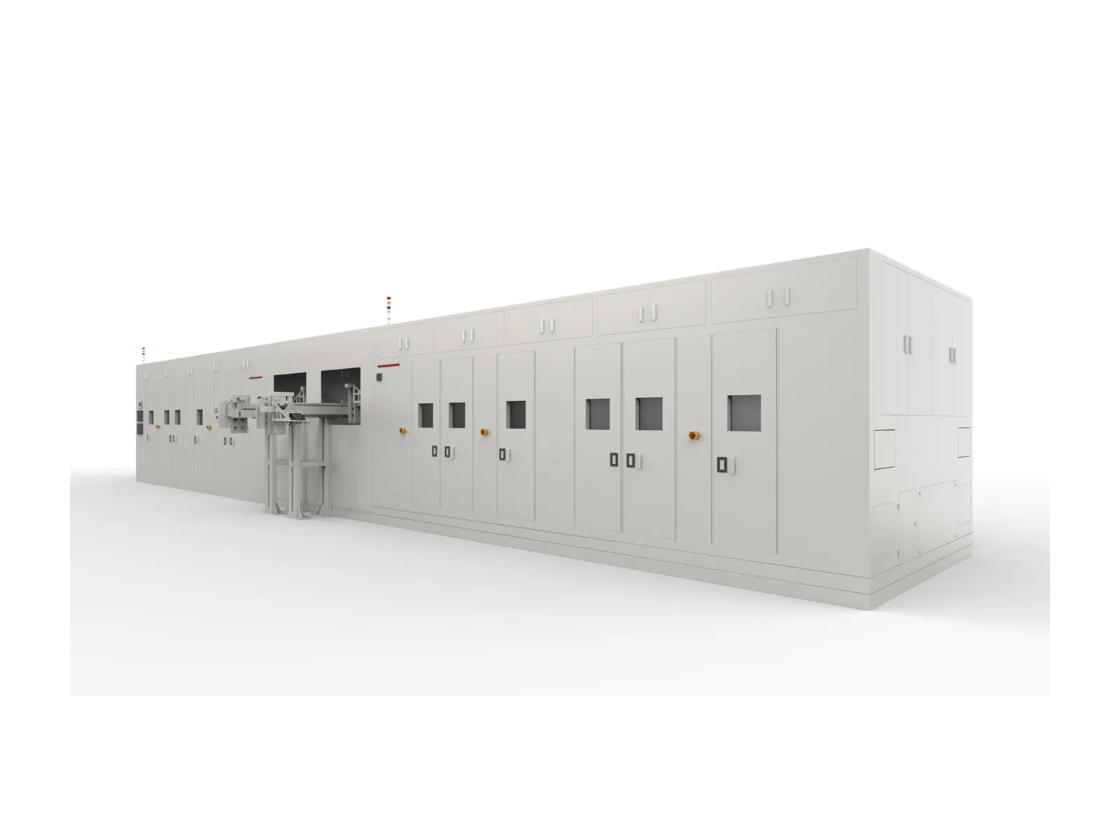
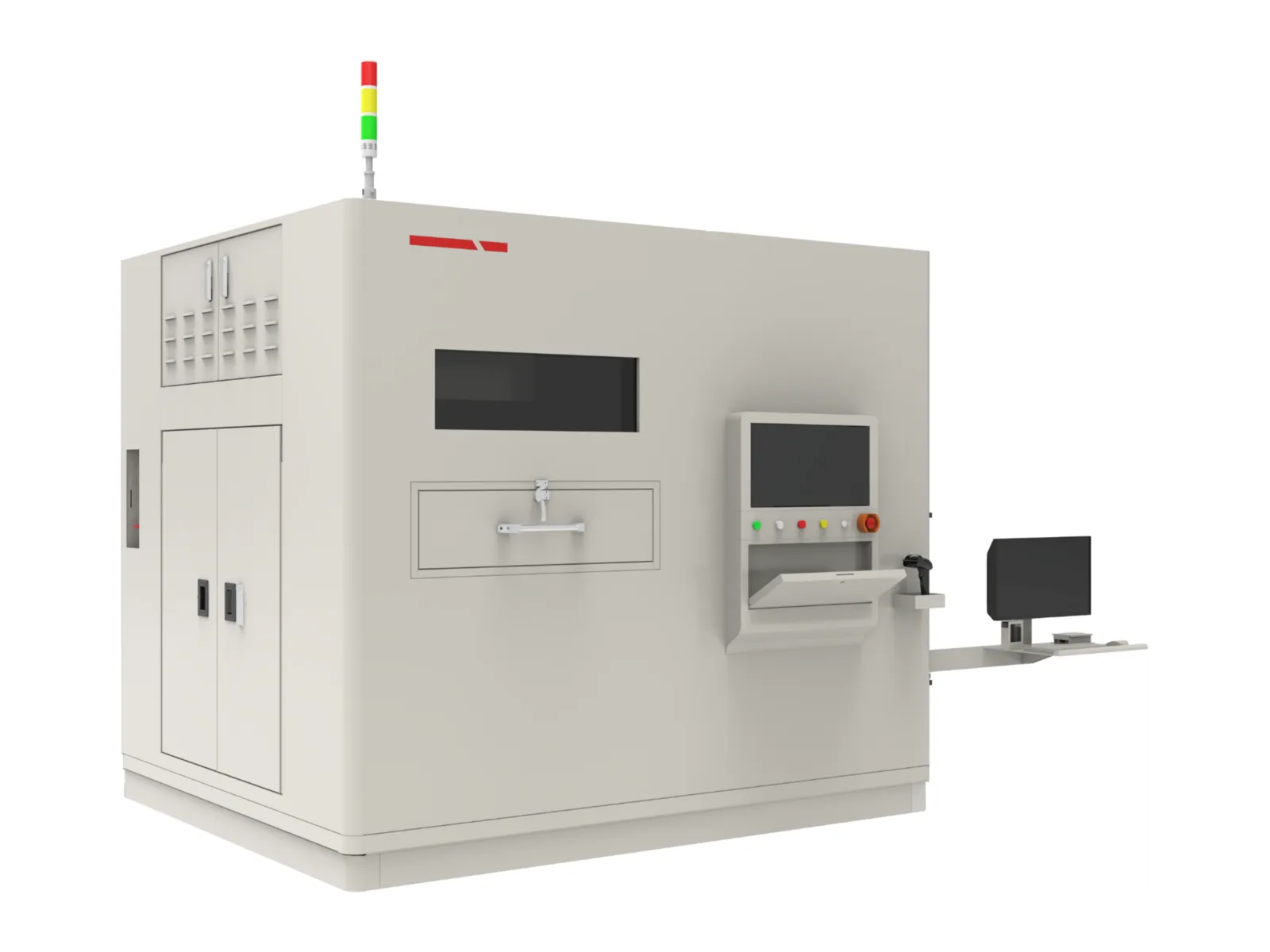
Royma offers X-ray and computed tomography (CT) for every stage of cell and module production. In the laboratory the RMCT4000 and RMCT6000 scan single cells, modules and their welds with microfocus and mini-focus sources; the desktop RMCT1000 handles coin cells, separators and small samples on the bench. Beside the line, the Quick CT checks the four corners of a prismatic cell for overhang and the tab welds in one cycle, loaded by hand and reporting a pass or fail result.
In the line, the RMLCT7500 inspects every prismatic cell in 3D at line speed: a corner scan takes 4 to 8 seconds, the resolution is below 30 µm, and the number of gantry rings sets the throughput, up to 36 cells per minute. The RMCLT1850 does the same for cylindrical and button cells, scanning several cells at once. For modules and packs the RMCT7000 gantry CT scans long parts lying flat, up to 2000 × 420 × 160 mm in the standard configuration, and the RMCT10000 penetrates complete packs with its linear accelerator source.
Every system delivers the same kind of result: the overhang of every layer, the position of every particle and the geometry of every weld, measured on the volume and written to the cell record. Cell makers gain a 100 % inspection where they had samples, and a laboratory result they can trust when a line result is questioned.
In practice
What the inspection returns.
The analysis software measures the anode-to-cathode overhang at every corner and every layer and compares it with the tolerance set for the cell type. Foreign particles are reported with size and position between the layers, so the risk of a separator puncture is judged from the data rather than from a suspicion. Weld results cover pore volume, melt depth and the seam position on the tab and the cover.
Inline results go to the line controller and the manufacturing execution system as a pass or fail decision with the measured values; laboratory results go into a report with slices, renderings and the measurement table. Royma supports the set-up for each cell type, from the scan recipe to the tolerance limits, and adds new cell formats as the line changes.
Questions
Questions about CT for batteries.
- Can CT inspect every cell in production, or only samples?
- Both. The RMLCT7500 and the RMCLT1850 are inline CT systems for 100 % inspection of prismatic, cylindrical and button cells at line speed. The Quick CT works at the line with manual loading, and the laboratory systems scan samples in full detail.
- What does 2D X-ray miss that CT shows?
- A 2D image overlays all electrode layers, so a small overhang fault on one layer hides behind the others. CT separates the layers and measures the overhang on each one, which is why the corner scan of a prismatic cell is done in 3D.
- Which system suits battery modules and packs?
- The RMCT7000 gantry CT scans long modules lying flat, with the part at rest while source and detector rotate. Complete packs with thick housings are scanned with the RMCT10000, whose linear accelerator source penetrates where conventional X-ray tubes stop.
- How are foreign particles reported?
- Each particle is located in three dimensions, with its size and its position relative to the separator and the electrodes. The result is written to the cell record, so a particle finding can be traced back to the cell and the production lot.
News
Application notes and news.
Application note ·
Why 2D X-ray misses prismatic-cell overhang faults – and what 100% inline 3D CT changes for LFP and ESS cell lines
Application note ·
Beyond the electrode stack: CT of top-cover laser welds, safety valves and seal balls on prismatic cells
Application note ·